
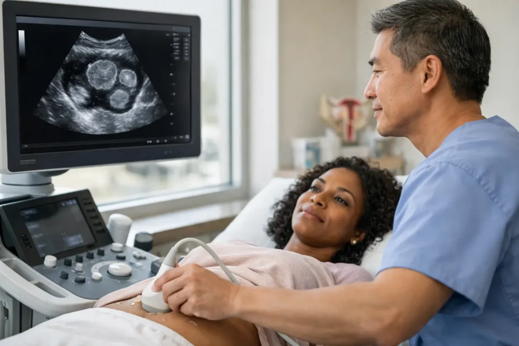
Heavy periods that interrupt work, pelvic pressure that will not settle, or unexpected difficulty conceiving can leave you looking for clear answers. So, can ultrasound detect fibroids? In many cases, yes. Pelvic ultrasound is usually the first imaging test used to look for fibroids, helping a qualified sonographer assess whether they are present, where they sit in the womb and how large they are.
Fibroids are very common, non-cancerous growths made of muscle and fibrous tissue. Some people never know they have them. For others, they can have a significant effect on comfort, bleeding, bladder and bowel symptoms, fertility, or pregnancy care. A scan cannot answer every question on its own, but it can be a valuable, reassuring first step towards the right care.
Can ultrasound detect fibroids accurately?
Ultrasound can detect many fibroids accurately, particularly when they are moderate or large in size. It uses sound waves to create real-time images of the uterus and surrounding pelvic structures, without radiation. During the examination, the sonographer can identify areas that look different from normal uterine muscle and take precise measurements.
The scan report may describe the number of fibroids seen, their dimensions and their position. This matters because location can influence symptoms and the next clinical conversation. A small fibroid on the outer surface of the womb may cause no concern at all, while one that changes the shape of the womb cavity may be more relevant for heavy bleeding or fertility.
Ultrasound is highly useful, but it is not infallible. Very small fibroids, fibroids in a difficult position, or a womb with several fibroids can sometimes be harder to assess in full. Body shape, bowel gas and the quality of the images can also affect what can be seen. If the results need further clarification, a clinician may recommend another type of assessment, such as MRI or hysteroscopy, depending on your symptoms and plans.
Which type of ultrasound is used for fibroids?
A pelvic ultrasound may be performed through the abdomen, internally, or as a combination of both. The most suitable approach depends on your symptoms, medical history and what needs to be assessed.
Transabdominal pelvic ultrasound
For a transabdominal scan, the probe moves gently over the lower tummy. You may be asked to arrive with a comfortably full bladder because this can improve the view of the uterus. This approach gives a broader picture of the pelvis and can be particularly helpful for larger fibroids that extend upwards from the womb.
Transvaginal pelvic ultrasound
A transvaginal scan uses a slim, covered probe placed gently into the vagina, with your consent. It often provides a closer, more detailed view of the womb lining, uterine muscle and ovaries. It can be especially useful for identifying smaller fibroids or assessing fibroids close to the uterine cavity.
You are always entitled to ask questions, request a chaperone and pause or stop an internal scan at any point. A compassionate clinical team should explain what is involved beforehand, protect your privacy and make sure you feel comfortable throughout.
What can a fibroid scan tell you?
A scan does more than simply confirm whether a fibroid is there. It helps build a clearer picture of what may be contributing to your symptoms. Fibroids are commonly described by where they grow:
- Intramural fibroids grow within the muscular wall of the womb.
- Subserosal fibroids grow towards the outside of the womb.
- Submucosal fibroids grow towards the inner cavity of the womb.
- Pedunculated fibroids are attached by a narrow stalk.
These descriptions may sound technical, but they help your doctor understand the practical implications. For example, submucosal fibroids can be more closely associated with heavy periods and may affect implantation, even when they are not especially large. Larger subserosal fibroids may be more likely to cause fullness, pressure or a frequent need to pass urine.
The scan may also assess the womb lining and ovaries, where appropriate. That is useful because heavy bleeding and pelvic pain can have more than one cause. Adenomyosis, ovarian cysts, endometrial polyps and some non-gynaecological conditions can produce overlapping symptoms. An expert scan helps distinguish between possibilities, although final diagnosis and treatment decisions should always consider your wider history.
Symptoms that may prompt a fibroid ultrasound
Not everyone with fibroids needs treatment, and not every heavy period is caused by a fibroid. However, arranging an assessment is sensible if symptoms are affecting your everyday life or you have concerns about your reproductive health.
Common reasons to seek advice include periods that are unusually heavy, prolonged or painful; bleeding between periods; pelvic pain or pressure; a bloated feeling; constipation; frequent urination; pain during sex; and difficulty becoming pregnant. Some people also discover fibroids during a routine pregnancy or fertility scan, having had no noticeable symptoms beforehand.
Seek urgent medical advice if you are soaking through pads or tampons very quickly, feel faint or short of breath with heavy bleeding, have severe sudden pelvic pain, or are pregnant and worried about pain or bleeding. Ultrasound can be helpful, but urgent symptoms should not wait for a routine appointment.
Fibroids and pregnancy: what does ultrasound show?
Finding a fibroid during pregnancy can feel alarming, especially when you were expecting the scan to focus only on your baby. In most pregnancies, fibroids do not prevent a healthy outcome. Their relevance depends on their size, number and position, as well as how they relate to the placenta, cervix and the space around the baby.
Pregnancy ultrasound can document fibroids and monitor them when clinically appropriate. Some fibroids may grow during pregnancy due to hormonal changes, while others remain stable or become less noticeable later on. Your maternity team will consider the findings alongside your symptoms and the progress of the pregnancy.
A fibroid may occasionally be linked with discomfort, particularly if it outgrows its blood supply and becomes painful. In some circumstances, fibroids can affect the baby’s position or influence birth planning. This does not mean complications are inevitable. It means care can be tailored with the right information and follow-up.
When ultrasound may not be the whole answer
If ultrasound findings do not fully explain your symptoms, further investigation may be appropriate. MRI can provide more detailed mapping of fibroids, particularly before certain treatments or surgery. Hysteroscopy involves looking inside the womb cavity and may be recommended when a fibroid appears to project into that space. Blood tests may also be advised if heavy periods raise concerns about iron deficiency or anaemia.
The best next step depends on what the scan shows and what matters most to you. Someone managing mild symptoms may choose monitoring and symptom relief. Someone with severe bleeding, pain, fertility concerns or pressure symptoms may need a gynaecology referral to discuss medicines, procedures or surgery. There is no one-size-fits-all treatment plan.
Preparing for your pelvic ultrasound
When you book, check whether you need a full bladder. This is commonly requested for a transabdominal pelvic scan and can make a meaningful difference to image quality. Wear comfortable clothing that allows easy access to your lower abdomen, and bring details of your cycle dates, symptoms, medications and any previous scan reports if you have them.
Try to describe symptoms as specifically as you can. Note whether bleeding has changed, where pain is felt, how long it lasts and whether it relates to your period, sex, eating or passing urine. These details help the clinician interpret the scan in context rather than looking at an image in isolation.
At Miracle Scan, diagnostic pelvic ultrasound is performed in a professional, supportive setting by qualified sonographers. You should leave with a clear understanding of the scan process and guidance on appropriate next steps, while recognising that ongoing symptoms or abnormal findings may need review by your GP or specialist.
A fibroid diagnosis can bring relief because it gives a name to symptoms that may have been difficult to live with. Whether your scan finds fibroids or points towards another explanation, clear imaging and compassionate clinical advice can help you move forward with greater confidence.